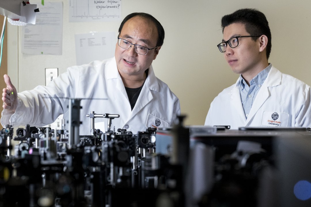
A team of scientists led by Oregon State chemist Chong Fang has discovered a new way to visualize and track chloride ions in living systems, opening the door to accelerated research on diseases like cystic fibrosis, epilepsy and certain cancers. Published in the journal Proceedings of the National Academy of Sciences, the breakthrough challenges longstanding assumptions about fluorescent protein biosensors and introduces a sharper, more reliable tool for studying chloride-related processes in the body.
Chloride is essential to life, playing a critical role in maintaining proper fluid balance within the body, aiding in digestion and supporting nerve and muscle function. But despite its importance, it remains difficult to study inside living systems. Tracking chloride requires biosensors, specialized protein-based tools that change their fluorescence when chloride is present. Most existing sensors become dimmer when chloride binds, making it hard to distinguish signals from background noise.
The new biosensor, called ChlorON3 does the opposite: it lights up. That “turn on” response gives scientists a sharper, more reliable tool for tracking chloride activity in real time.
“We now have a powerful biosensor that is non-toxic, genetically encodable and can detect the ‘queen of electrolytes,’” said Fang, referring to chloride, the second most abundant electrolyte in the human body after sodium. Fang is a professor in the Department of Chemistry and currently holds the Patricia Valian Reser Endowed Faculty Scholar position.
What makes this biosensor shine is something no one expected. Using ultrafast laser spectroscopy with the resolution of a few millionth of a billionth of a second and advanced computer simulations, the team found that the heart of the biosensor, a component called a chromophore, becomes twisted and rigid when chloride is present.
These findings overturn the conventional belief that chromophores need to be flat, or planar, to glow brightly. Instead, the research shows that rigidity, not planarity, is the decisive factor in producing a strong fluorescent signal.


